
 +86-0755-86131192
 2026-08-19
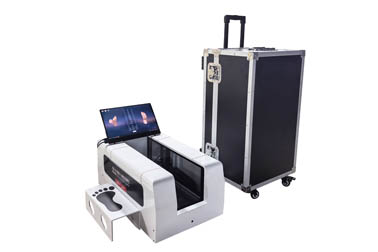
The 3D laser foot scanner is a device that adopts non‑contact laser scanning technology to rapidly construct high‑precision three‑dimensional foot models, and obtain the morphological structures and relevant geometric parameters of human feet under weight‑bearing or non‑weight‑bearing conditions in real time.
Flat feet and high arches are common foot morphological abnormalities, marked by noticeable changes in arch height. Obvious deviations occur in the force‑bearing structure and physical appearance of feet when people stand or walk.
The 3D laser foot scanner can measure objective data such as foot volume, surface area, arch height, foot width and ankle joint angle under different conditions. Featuring high‑resolution data and morphological visualization, it enables more intuitive and accurate assessment of foot conditions including flat feet and high arches.
When conducting foot assessments, the 3D laser foot scanner generally supports two scanning modes: static weight‑bearing scanning and static non‑weight‑bearing scanning.
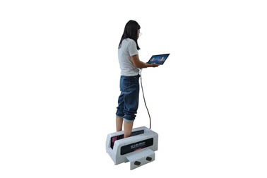
For static weight‑bearing scanning, subjects simply stand barefoot on the scanning platform, keep the body upright, place both feet naturally apart, and distribute body weight evenly. No physical contact with equipment is required throughout the process, ensuring excellent hygiene. Scanning is completed in a very short time, usually within 5‑10 seconds.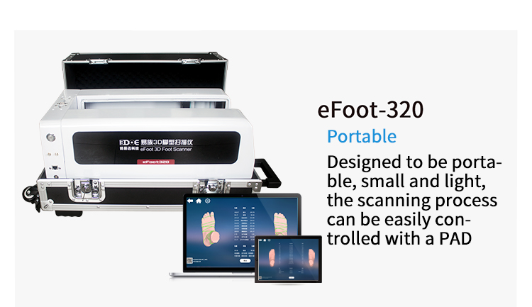
Static weight‑bearing scanning mainly reconstructs the three‑dimensional morphology of feet under load. It is suitable for rapid screening and file establishment of foot appearance deformities.
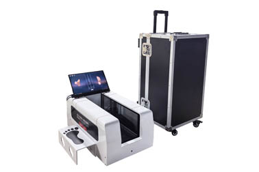
In static non‑weight‑bearing scanning, the subject’s feet are suspended or free from gravitational compression. The device captures the native three‑dimensional structure of feet in a naturally relaxed state, and fully restores the original morphology of bones and soft tissues, including natural arch curvature, instep height, foot girth data, volume distribution of each region, comparison between left and right feet, heel tilt angle and other parameters.
High arches or flat feet are often accompanied by changes in foot volume and morphology. Measuring morphological data delivers extremely high assessment value and enables more three‑dimensional evaluation of foot structures.
A common indicator for evaluating arch morphology is the Arch Height Index (AHI), a core morphological metric.
Arch Height Index (AHI) = Vertical height of the lowest arch point ÷ Effective foot length
A lower Arch Height Index value indicates a lower vertical arch height and a larger plantar contact area, which means a more severe degree of flat feet. A higher value represents a more raised foot arch and more suspended plantar area, pointing to more prominent high‑arch deformity.
Nevertheless, since the foot bones of adolescents have not yet closed, conclusions cannot be drawn solely based on a single scanning result. Comprehensive judgment shall be made in combination with growth‑development curves and dynamic gait data of plantar pressure.
Advantages of foot assessment using the 3D laser foot scanner:
1. Strong data visualization. The generated 3D model can be rotated for 360‑degree observation. It avoids the mess and discomfort brought by traditional plaster casting, and is especially suitable for scenarios with high hygiene requirements, such as high‑end footwear customization and medical rehabilitation centers.
2. Diverse measurement dimensions. It can acquire not only planar area but also volume, height and girth. Some high‑end models support multimodal analysis combined with pressure data, enabling assessment and judgment from both geometric and mechanical perspectives for more three‑dimensional and comprehensive evaluation.
3. Wide‑ranging applications. The acquired three‑dimensional data serves as the technical foundation not only for screening flat feet and high arches, but also for digital orthotic insole design, sports shoe last development, pre‑operative and post‑operative morphological comparison, foot growth and development monitoring, virtual shoe‑fitting and other technologies.
However, the morphological data measured by the 3D laser foot scanner mainly reflects external structures and cannot fully represent functional status.
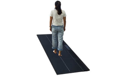
As it mainly presents geometric shapes under “static” or “quasi‑static” conditions, it cannot directly reflect muscle activation during walking. Physicians need to make comprehensive diagnoses by combining dynamic gait analysis and clinical physical examination。
 Back to list


 +86-0755-86131192